|
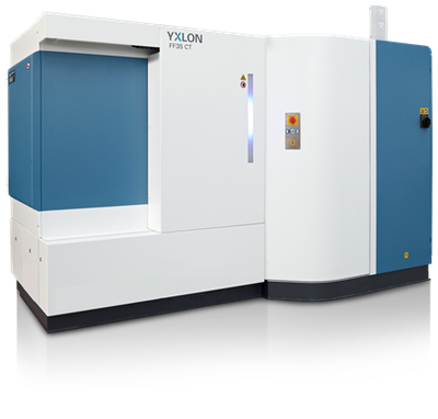
Since December 2020, the NHM Vienna will have a micro-computed tomography (micro-CT) device. It enables non-destructive insights into a wide variety of organisms and objects and allows the determination of the spatial distribution of matter inside a sample as well as the generation of three-dimensional volume and surface models of objects with sizes from the sub-millimeter range (e.g. insect eyes) to a size of several decimeters (e.g. pick handle). The device is suitable for the acquisition of 2D X-ray images as well as 3D computed tomography.
The micro-CT is assigned to the central research laboratories of the NHM Vienna and can be used by every department of the museum. External users are also welcome - whether within a national or international academic research cooperation or as a commercial user on a contractual basis.
The device
The YXLON FF35 CT is equipped with a YXLON 225 kV micro-focus directional beam tube and a YXLON 190 kV nano-focus transmission tube, which can be operated in two or four different power levels. Two different targets (tungsten and diamond) are available for beam generation. In this way, both millimeter-sized samples with high resolution and centimeter to decimeter-sized samples can be analyzed with high performance. The detector system from YXLON (Y.Panel 4343 CT CsJ, 2880 x 2880 pixels, pixel pitch 150 µm) offers the option of expanding the measuring field.

Specifications
- Maximum CT measuring range: approx. 510 mm Ø x 600 mm height
- Sample space: 530 mm Ø x 800 mm height (collision protection through manually defined cylinder); 300 mm x 500 mm high (collision protection through automated shape recognition "IntelliGuard" via camera)
- Max. Sample weight: 30 kg
- Focus-detector distance: Micro-focus: 508 - 1175 mm; Nano-focus: 448 - 1115 mm
- Focus-object distance: up to 965 mm
X-ray tubes:
1. YXLON FXT 225.48 micro-focus directional beam tube up to 225 kV water-cooled; up to 280 W at the target or 320 W tube power. Detectability of details in the fluoroscopic image of less than 3 μm
2. YXLON FXT 190.61 open nano-focus transmission X-ray tube from 20 kV to 190 kV water-cooled; up to 15 W at the target or 80 W tube power. Detection of details in the fluoroscopic image of less than 200 nm
Detector: Digital flat detector Y.Panel 4343 CT CsJ, detector size 43 x 43 cm, pixel pitch 150 μm, pixel matrix 2880 x 2880 (1x1) / 1440 x 1440 (2x2), frame rate up to 15 Hz (1x1) / 30 Hz (2x2) , 16 bit, scintillator CsJ.
Manipulator: Granite-based manipulator with vibration damping as well as length and angle measuring devices, software-supported automatic coordinate calibration
Air conditioning: cabin room temperatures acc. Measuring room quality class 3 according to VDI 2627 (temperature change in 1 hour max. 1.0 K and in 24 hours max. 2.0 K).
Filter and collimator set: pre-filters made of Al (0.5 / 1.0 / 2.0 mm), Cu (0.2 / 1.0 / 2.0 mm) and Sn (1.0 / 1.5 / 2.0 mm)
|
